Scanning cavity microscopy
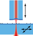
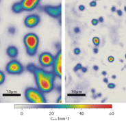
 Improving the sensitivity of optical microscopy could give access to the optical properties of a large class of interesting nanoscale systems on a single particle level. To boost sensitivity, we are developing a novel scanning microscopy technique that harnesses multiple interactions of probe light with an object. This is realized by placing the sample inside a high-Finesse optical microcavity, which provides a signal enhancement on the order of the cavity Finesse. Raster-scanning the sample through the cavity mode enables us to obtain spatial maps of sample absorption and dispersion of individual nanoparticles. We have developed a scheme to increase the spatial resolution of the images by combining the signal of higher-order transverse cavity modes.

The method should enable e.g. the spectroscopic characterization of individual nanoparticles, dispersive bio-sensing with high spatial resolution, and the detection of non-fluorescent single molecules.
More details can be found in our article
[Mader et al., Nature Communications 6, 7249 (2015)].
The project is partially funded by NIM.
